
Executive Summary
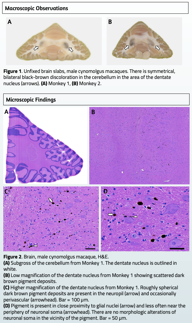
This poster documents an incidental finding: melanin pigment inside the cerebellar dentate nucleus of two young cynomolgus macaques.
Melanin shows up elsewhere in primate brains as a normal feature, in the meninges, cortex, substantia nigra, and locus coeruleus, but not typically in the dentate nucleus. The same pattern has been seen in people. As far as the authors know, this is the first time it’s been reported in a non-human primate, which makes the case worth flagging for anyone reviewing pigmented lesions in NHP neuropathology.
Table of Contents
Add a header to begin generating the table of contents
Introduction
- Pigment in the central nervous system (CNS)
- In cynomolgus macaques, melanin pigment occasionally occurs in the meninges, as a product of leptomeningeal melanocytes, and in the cerebral cortex, where it is often perivascular and associated with astrocytes.
- Neuromelanin is a type of melanin pigment produced in catecholaminergic neurons, where it accumulates in the neuronal cytoplasm with age, most notably in the substantia nigra and locus ceruleus.
- Non-melanin pigments, such as hemosiderin and lipofuscin, can also occur in the CNS.
- Melanosis of the dentate nucleus (DN) is a rarely reported condition in people.
- Between 1941-2017, there were 19 reported cases (13 females, 6 males) ranging in age from 26-104 years (mean 70.2, median 72).
- There are no common clinical conditions or histories among affected individuals.
- Pigment in the DN has not been previously reported in non-human primates.
Methodology
- Both affected animals were 18-month-old male Chinese origin cynomolgus macaques
- A single dose of an AAV gene therapy was administered direct to the brain in a region remote from the cerebellum
- On study, the animals had regular detailed clinical observations. Neurologic assessments were also performed.
- Euthanasia occurred at scheduled study termination, 58 days post dose.
- At necropsy, animals were perfused with saline. The unfixed brains were coronally sectioned followed by immersion fixation in 4% paraformaldehyde.
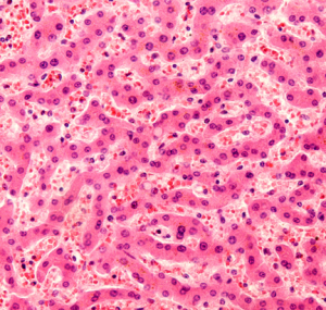
- Fixed brain samples were routinely processed for paraffin embedding and sectioning. Tissue sections were stained with hematoxylin-and-eosin (H&E).
Results
- Both animals were euthanized at scheduled study termination. There were no notable in life findings for either animal.
- The pigment did not autofluoresce.
- Pigment deposits were not identified elsewhere in the examined brain or spinal cord of these animals
Conclusions
- This finding was not associated with any neurologic abnormalities.
- No relationship to the test article was determined in this study.
- This may represent a rare spontaneous condition in cynomolgus macaques.
- The location and microscopic appearance of the pigment in these two macaques closely resembles cases of melanosis of the dentate nucleus reported in people.
- The underlying genesis and clinical significance remains unknown.
- Melanin in the DN has been demonstrated within astrocytic processes.
- Melanin deposits in the cerebral cortex in macaques have also been associated with astrocytes suggesting a potential shared mechanism of formation.
- A process of involution and/or a relationship to lipofuscin has been postulated for melanosis of the DN, based on the generally advanced age of affected individuals. However, the young age of these affected cynomolgus monkeys suggests other factors, perhaps even congenital factors, play a role in this condition.
- Identification of additional cases of melanosis of the DN may shed further light on the pathogenesis of this rare condition in both cynomolgus macaques and humans.