


Executive Summary
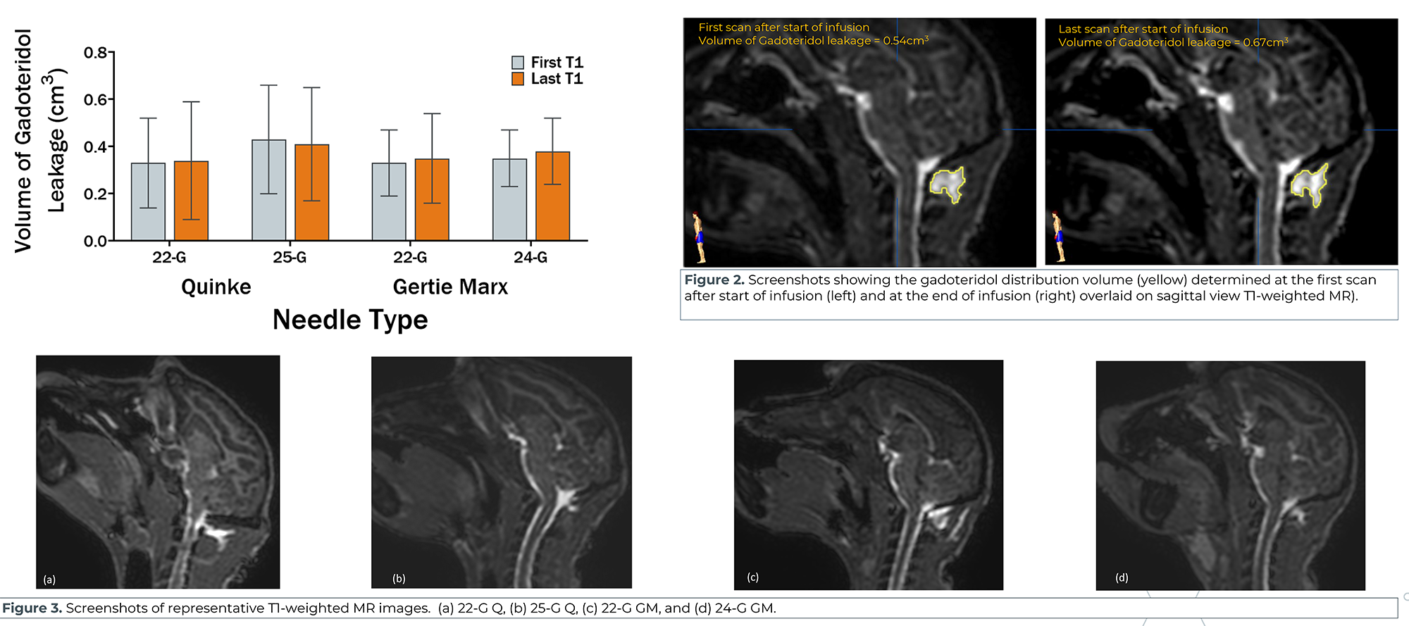
This research compares needle types used for cisterna magna dosing in cynomolgus monkeys, checking whether needle style or gauge affects cerebrospinal fluid leakage. Five animals received doses through Quinke and Gertie Marx needles at different gauges, with MRI used to track gadolinium distribution outside the CSF space.
No needle type or gauge showed a clear leakage advantage over the others.
The study points to needle position at the dura and small movements during the procedure as the more likely sources of leakage. That shifts the emphasis toward operator technique rather than hardware choice.
Introduction
Central delivery into the cisterna magna (CM) via percutaneous administration with spinal needles is vital for a variety of therapeutics tested in non-human primates (NHP). Correct administration can be affected by multiple factors: animal position, experience and technique of the dose administrator, needle type (e.g., Cutting, pencil point), needle gauge, cerebrospinal fluid (CSF) collection, or volume of therapeutic. Cerebrospinal fluid leakage during and after dose administration is a concern. The effect that the type and gauge of spinal needle had on leakage was evaluated using magnetic resonance imaging.
Methods
Five cynomolgus NHPs were anesthetized and prepared for CM dose administration. Induction was performed using ketamine (5mg/kg), dexmedetomidine (0.025mg/kg), and buprenorphine (0.02mg/kg). Anesthesia was maintained with a mixture of oxygen (1L/min) and isoflurane (1-3%). Buprenorphine ER (0.2mg/kg) and Meloxicam ER (0.6mg/kg) were administered in recovery.
22-G Quinke (Q), 25-G Q, 22-G Gertie Marx (GM) and 24-G GM spinal needles were evaluated. Selection of needle type and gauge varied between animals on dosing days. Dose administrator and holder remained consistent throughout the study to reduce variability. 1.0 mL of CSF was collected immediately prior to administration of 2.0 mL of 2mM Prohance (gadoteridol) in Phosphate Buffered Saline. Immediately following administration, T1-weighted and FLAIR scans were obtained with the MRI (Philips Achieva Quasar 3.0T MR). Continuous scanning occurred for ~30 minutes. The animals were recovered and allowed a two-week washout period prior to the next dose/needle type. Gadoteridol distribution volume outside the CSF space was considered as CSF leakage and was quantified by Brainlab (Brainlab AG, Munich, Germany). For volumetric assessment, a customized algorithm for segmentation of the gadoteridol distribution by subtracting the baseline images from the first and the last post infusion T1-weighted MR images was applied. (Figure 2).
Results