
Introduction
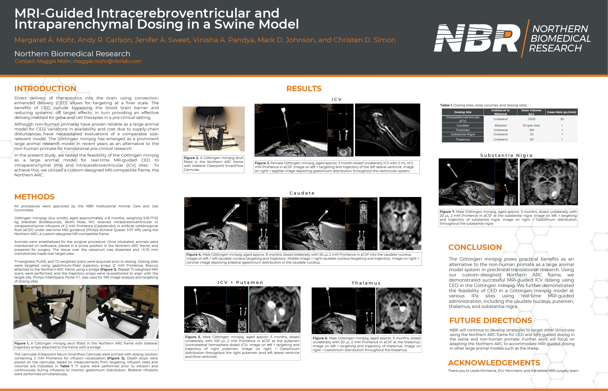
Direct delivery of therapeutics into the brain using convection-enhanced delivery (CED) allows for targeting at a finer scale. The benefits of CED include bypassing the blood brain barrier and reducing systemic off target effects, in turn providing an effective delivery method for gene and cell therapies in a pre-clinical setting.
Although non-human primates have proven reliable as a large animal model for CED, variations in availability and cost due to supply-chain disturbances have necessitated evaluations of a comparable size relevant model. The Göttingen minipig has emerged as a prominent large animal research model in recent years as an alternative to the non-human primate for translational pre-clinical research.
In the present study, we tested the feasibility of the Göttingen minipig as a large animal model for real-time MR-guided CED to intraparenchymal (IPa) and intracerebroventricular (ICV) sites. To achieve this, we utilized a custom-designed MR-compatible frame, the Northern ARC.
Methods
All procedures were approved by the NBR Institutional Animal Care and Use Committee.
Göttingen minipigs (Sus scrofa), aged approximately 4-8 months, weighing 9.16-17.92 kg (Marshall BioResources, North Rose, NY) received intracerebroventricular or intraparenchymal infusions of 2 mM ProHance (Gadoteridol) in artificial cerebrospinal fluid (aCSF) under real-time MRI guidance (Philips Achieva Quasar 3.0T MR) using the Northern-ARC, a custom-designed MR-compatible frame.
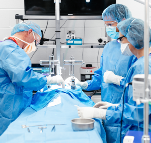
Animals were anesthetized for the surgical procedure. Once intubated, animals were maintained on isoflurane, placed in a prone position in the Northern ARC frame, and prepared for surgery. The tissue over the calvarium was dissected and ~5-10 mm craniotomies made over target sites.
T1-weighted, FLAIR, and T2-weighted scans were acquired prior to dosing. Dosing sites were targeted using gadolinium-filled trajectory arrays (2 mM ProHance, Bracco) attached to the Northern ARC frame using a bridge (Figure 1). Repeat T1-weighted MRI scans were performed, and the trajectory arrays were re-positioned to align with the target site. Philips IntelliSpace Portal 11.1. was used for MR image analysis and targeting of dosing sites.
The cannulae (Clearpoint Neuro Smartflow Cannula) were primed with dosing solution containing 2 mM ProHance for infusion visualization (Figure 2). Depth stops were placed on the cannulae, based on measurements from targeting. Infusion rates and volumes are indicated in Table 1. T1 scans were performed prior to infusion and continuously during infusions to monitor gadolinium distribution. Bilateral infusions were performed simultaneously.
Results
Click on poster below.
Conclusion
The Göttingen minipig poses practical benefits as an alternative to the non-human primate as a large animal model system in preclinical translational research. Using our custom-designed Northern ARC frame, we demonstrated successful MRI-guided ICV dosing using CED in the Göttingen minipig. We further demonstrated the feasibility of CED in a Göttingen minipig model at various IPa sites using real-time MRI-guided administration, including the caudate nucleus, putamen, thalamus, and substantia nigra.
Future Directions
NBR will continue to develop strategies to target other structures using the Northern ARC frame for CED and MRI-guided dosing in the swine and non-human primate. Further work will focus on adapting the Northern ARC to accommodate MRI-guided dosing in other large animal models such as the sheep.
Acknowledgements
Thank you to Leslie Minnema, Eric Herrmann, and the entire NBR surgery team.